

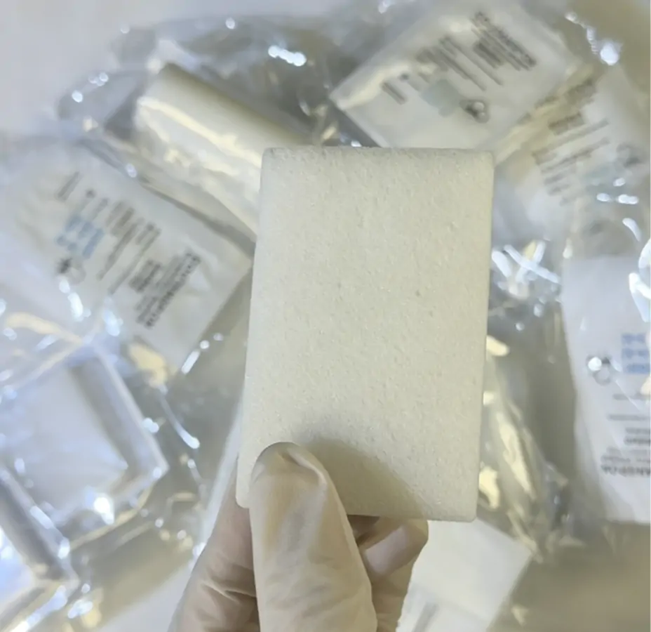
Why Investing in High-Quality Hemostatic Gelatin Sponges Reduces Surgical Complications and Optimizes Hospital Supply Chains
1. The Cellular Mechanism of Absorbable Gelatin Sponge Hemostasis
When dealing with capillary or venous bleeding where traditional ligation is impractical, an absorbable gelatin sponge provides a critical structural matrix. Upon contact with bleeding tissue, the porous matrix of the sponge absorbs roughly forty times its weight in blood and fluid. This rapid absorption causes the physical matrix to swell, trapping platelets within its interstitial spaces.
Unlike chemical clotting agents, the primary action here is mechanical. The damaged platelets disintegrate, releasing thromboplastin, which accelerates the natural coagulation cascade. In general surgery and orthopedic cases, achieving this mechanical seal quickly reduces the total volume of blood loss and minimizes the need for prolonged manual compression. The biological compatibility of purified porcine skin gelatin ensures that the material triggers minimal inflammatory response while providing a stable framework for the initial clot. Within four to six weeks, the entire sponge is completely liquefied and absorbed by the surrounding tissues without leaving a permanent foreign body.
2. Clinical Scenarios Across Diverse Surgical Disciplines
In the operating room, surgical teams encounter distinct challenges depending on the vascularity of the targeted organ. During laminectomies or deep orthopedic procedures, oozing from cancellous bone can obscure the visual field and delay the placement of hardware. Here, the sponge is frequently packed tightly into bony crevices to tamponade the bleeding.
In ENT procedures, particularly after a functional endoscopic sinus surgery, a slightly modified, compressed version of the sponge is placed in the nasal cavity to manage mucosal bleeding while maintaining airway patency. For general surgeons performing hepatic resections, the sponge is often moistened with sterile saline or a topical thrombin solution and applied directly to the raw liver surface. In each of these settings, the material adapts to irregular anatomical contours, providing a versatile solution that transitions easily from a dry, firm compress to a soft, compliant packing material that adheres securely to the bleeding site.
3. Comparing Standard Alternatives: Gelatin Sponge vs Gelfoam
Hospital clinical evaluation committees routinely compare brand-name standards with specialized market alternatives to balance clinical efficacy and budget targets. When looking at gelatin sponge vs gelfoam, the primary clinical variables center around porosity uniformity, hydration speed, and structural integrity under pressure. While Gelfoam represents the historical benchmark for predictability, modern advanced alternatives often feature enhanced tensile strength when wet.
Standard options can sometimes become friable or tear when saturated with blood, which complicates handling with surgical forceps and may require multiple reapplications. Newer alternatives utilize cross-linking manufacturing techniques that preserve the sponge’s shape even when handled roughly during deep pelvic or spinal surgeries. This prevents the material from fragmenting prematurely, ensuring that the entire clot matrix remains intact at the wound site rather than breaking apart and leaving microscopic remnants that could theoretically increase the risk of localized granuloma formation.
4. Procurement Metrics and Supply Chain Cost Dynamics
From a hospital procurement perspective, surgical hemostasis materials represent a significant portion of the recurring disposable medical consumables budget. Choosing the right supplier involves looking past the initial unit cost to analyze the total cost of care. When a surgeon uses a lower-grade sponge that tears easily, they frequently open a second or third blister pack to achieve adequate hemostasis, which drives up the per-procedure material cost.
Furthermore, premium sponges that offer rapid hydration reduce the total time spent waiting for hemostasis during delicate steps. This directly translates into lower operating room utilization costs, which can average tens of dollars per minute. By standardizing high-performing alternative gelatin sponges that match the clinical metrics of legacy brands, healthcare networks can consolidate their vendor lists, negotiate better volume discounts, and reduce inventory overhead without compromising patient safety or surgeon satisfaction.
5. Infection Control and Mucosal Safety Protocols
Patient safety guidelines emphasize that any implantable biomaterial must strictly adhere to rigorous biocompatibility standards to avoid postoperative complications. Modern absorbable gelatin sponge manufacturing involves stringent purification processes to eliminate cellular debris and pyrogens from the raw porcine collagen source. This focus on purity minimizes the risk of a foreign-body reaction, which is crucial when the material is placed near delicate structures like cranial nerves or sensitive mucosal tissue.
In rhinological applications, maintaining mucosal safety means the sponge must not cause dense adhesion formation or long-term mucosal irritation, which could impair the natural mucociliary clearance of the sinuses. Additionally, proper gamma irradiation sterilization ensures that the product arrives completely sterile, eliminating risks of bacterial contamination that could lead to deep-site infections in compromised or immunocompromised surgical patients.
6. Postoperative Care and Clinical Guideline Compliance
International surgical guidelines, including recommendations from the World Health Organization and the American College of Surgeons, place a strong emphasis on meticulous local hemostasis to prevent postoperative hematoma formation. A postoperative hematoma can cause tissue ischemia, delay wound healing, and require an unplanned return to the operating room. Using an absorbable gelatin sponge helps ensure that the surgical site remains stable during the critical first twenty-four to forty-eight hours after closure.
Because the material liquefies naturally, it does not require a painful physical removal process, which is especially beneficial in pediatric cases or sensitive ENT procedures. Nurses monitoring patients postoperatively report fewer dressing changes and lower pain scores when internal oozing is controlled effectively at the source, streamlining the clinical workflow and allowing for faster patient discharge from the post-anesthesia care unit.
📚 References
- World Health Organization. (2016). Global Guidelines for the Prevention of Surgical Site Infection. World Health Organization.
- Schonauer, C., Tessitore, E., Barbagallo, G., Albanese, V., & Moraci, A. (2004). The use of local hemostatic agents in neurosurgery: a review. European Spine Journal, 13(1), S89-S96.
- Acharya, S., & Bareja, R. (2020). Comparative evaluation of various topical hemostatic agents in surgical practices. Journal of Clinical and Diagnostic Research, 14(8), MC01-MC05.