

Why Standardizing on a High-Quality Sterile Ultrasound Probe Cover Optimizes Patient Outcomes and Reduces Hospital Procurement Overhead
1. Defining the Protective Role of an Ultrasound Probe Cover in Contemporary Infection Control
In the fast-paced ecosystem of modern hospitals, diagnostic imaging is no longer confined to the radiology department. Point-of-care ultrasound (POCUS) is heavily utilized across emergency rooms, intensive care units, and interventional suites. While these devices provide invaluable real-time visualization, the transducer itself acts as a potential vector for cross-contamination. An ultrasound probe cover serves as the primary mechanical barrier designed to isolate the patient’s skin or mucosal membranes from the acoustic lens of the equipment. Relying solely on liquid chemical disinfection between patient consultations can fall short, particularly when dealing with non-intact skin or asymptomatic carriers of highly resilient pathogens. By integrating a dedicated material barrier into standard protocols, healthcare facilities can maintain strict compliance with international safety standards while protecting complex, delicate transducer arrays from the structural degradation associated with aggressive chemical wiping.
2. The Clinical Necessity of an Absolute Sterile Ultrasound Probe Cover in Interventional Procedures
When a diagnostic procedure transitions into an invasive intervention, the regulatory and hygienic requirements for equipment preparation escalate dramatically. Utilizing a sterile ultrasound probe cover becomes an absolute necessity during regional anesthesia, central venous line placements, and intraoperative scanning. In these clinical scenarios, any breach in the sterile field directly threatens patient safety, potentially leading to deep tissue infections or healthcare-associated bacteremia. Clinical evidence suggests that standard non-sterile sheaths or makeshift wraps do not provide adequate protection against microscopic pathogens. Choosing a verified, medical-grade barrier ensures that the device can safely enter critical surgical zones. For hospital procurement teams, evaluating the physical integrity, viral penetration resistance, and tear strength of these consumables is crucial. Standardizing on validated options helps risk management departments lower institutional liability and protect vulnerable populations from preventable cross-contamination.
3. Addressing the Vulnerabilities of Mucosal Barriers with a Dedicated Transvaginal Ultrasound Probe Cover
Specialized clinical environments require tailored barrier solutions, particularly in the fields of obstetrics and gynecology. Conducting pelvic examinations involves prolonged contact with sensitive mucosal tissue, which possesses a much lower natural defense mechanism against viral and bacterial transmission than intact skin. Implementing a specialized transvaginal ultrasound probe cover is essential for ensuring patient safety during these routine yet intimate diagnostic procedures. Mucosal layers are highly susceptible to micro-abrasions, meaning that any pathogen remaining on an improperly prepared transducer can easily establish an infection. Industry guidelines from organizations like the World Health Organization (WHO) emphasize that cross-contamination during endocavity scanning is a serious operational risk. Purchasing departments must prioritize procurement of sheaths specifically shaped and validated for endocavity applications to guarantee a precise fit, minimizing the risk of slippage or structural failure during the clinical examination.
4. Material Disadvantages and Safety Risks of Utilizing a Condom Ultrasound Probe Cover for Medical Imaging
In some clinical settings, a lack of immediate inventory or a desire to minimize upfront costs leads to the controversial practice of substituting medical sheaths with commercial alternatives. Using a standard latex condom ultrasound probe cover is a practice that introduces significant material and clinical disadvantages. Commercial condoms are engineered for a entirely different purpose and frequently contain added lubricants, fragrances, or spermicides that can chemically react with the sensitive acoustic lens materials of expensive ultrasound machines. Over time, these chemical additives can cause the matching layer of the transducer to swell, crack, or degrade, resulting in permanent image deterioration and premature equipment failure. Furthermore, commercial options often exhibit higher rates of micro-perforations under mechanical stress compared to dedicated medical-grade sheaths, and they pose a substantial risk for undetected latex allergies among patient populations and nursing staff alike.
5. Optimizing Workflow Efficiency: How to Put on Ultrasound Probe Cover Sheaths without Trapping Air
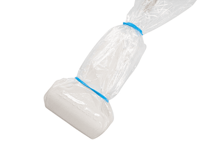
Achieving diagnostic accuracy while maintaining a sterile field requires precise handling during the pre-procedural preparation stage. Mastering how to put on ultrasound probe cover sheaths correctly is a fundamental operational skill for sonographers and interventional nurses. The workflow begins with a non-sterile assistant applying an adequate amount of acoustic coupling gel directly onto the active face of the transducer. It is vital to ensure that no air bubbles are trapped within this initial gel layer, as air gaps create severe acoustic artifacts that block the ultrasound waves, leading to distorted images and potentially compromised diagnoses. Next, the sterile operator carefully unrolls the protective sleeve over the probe assembly, pulling the material taut across the acoustic window. The sheath is then secured firmly in place using the medical-grade elastic bands provided in the kit, preventing the material from shifting during active scanning and allowing the clinician to focus entirely on the procedural target.
6. Supply Chain Standardization and the Economic Value of Quality Transducer Sheaths
Evaluating the financial trajectory of medical consumables requires a broader perspective than simply comparing unit purchase prices. When a hospital system standardizes its inventory on a dependable, multi-departmental line of imaging covers, it achieves significant operational efficiencies. Grouping orders reduces the administrative costs associated with managing multiple vendor contracts and prevents the waste generated by holding niche, short-expiry stock in separate departments. Furthermore, utilizing high-quality polyurethane or advanced latex-free polymers drastically extends the operational lifespan of capital imaging assets. By preventing fluids from entering the internal housing of the transducer and reducing the frequency of harsh chemical immersion, a reliable physical barrier preserves image clarity and prevents electronic component failure. Ultimately, investing in specialized, clinically proven sheaths lowers the total cost of ownership for diagnostic systems while maintaining an uncompromised standard of patient care.
📚 References
- World Health Organization (WHO): Decontamination and Reprocessing of Medical Devices for Health-care Facilities, Section on Ultrasound Transducer Hygiene and Endocavity Safety.
- Centers for Disease Control and Prevention (CDC): Guideline for Disinfection and Sterilization in Healthcare Facilities, Medical Device Classification and Semi-critical Barriers.
- American Institute of Ultrasound in Medicine (AIUM): Official Guidelines for Cleaning and Preparing External and Internal Ultrasound Transducers and Equipment between Patients.
- PubMed / Journal of Clinical Ultrasound: Comparative Analysis of Material Integrity and Acoustic Impedance: Medical-Grade Polyurethane Sheaths versus Commercial Latex Alternatives.